Open-source
Informatics Tools for Radiotherapy Research
This is the companion website to the book chapter
"Open-source Informatics for Radiotherapy Research," by Joseph
Deasy and Aditya Apte, in the book "Informatics in Radiation
Oncology," ed. George Starkschall and Alfredo ('Al') Siochi, CRC
Press (2013)
Last updated: October, 2013, by Joe Deasy, PhD.
(deasyj mskcc org ; you know what to do). We
intend to update it at some unspecified frequency. If you
have suggested improvements, additions, or corrections, please
email me.
This page lists radiotherapy-specific informatics tools
and resources and a few more general image handling and
statistical modeling tools. General DICOM tools are
referenced below, but are not the focus.
No fitness is implied for any of these tools for
treatment-related data processing or decision making.
Many of these tools are open-source and have widely
varying quality. This list is not meant to be
comprehensive.
DICOM-RT introduction pages
What is DICOM? Here is a nice presence courtesy of Elekta:
http://www.elekta.com/healthcare_international_what_dicom.php
Introductory course given by the Swiss Society of Radiobiology and
Medical Physics (SGMP). Informative slide sets are provided.
http://www.sgsmp.ch/sem03a-e.htm
Last updated: July 2003.
DICOM for RT introduction by Andrew Reilly. This pdf slide
show
gives examples of DICOM-RT troubleshooting as well as a list of
useful
tools (most are relisted here).
http://www.oncphys.ed.ac.uk/downloads/confs/DICOMRT.pdf
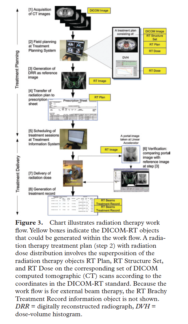
Law, M. Y. and B. Liu (2009). "Informatics in radiology: DICOM-RT
and
its utilization in radiation therapy." Radiographics 29(3): 655-667.
This is a useful short paper discussing the basics of
Radiation
Oncology information flow and DICOM-RT.
http://radiographics.rsna.org/content/29/3/655.short.
A figure from the paper:
General DICOM resource pages
A useful, brief, introduction to DICOM, as well as useful links, can
be
found at http://www.cabiatl.com/mricro/dicom/index.html.
"A single DICOM file contains both a header (which stores
information
about the patient's name, the type of scan, image dimensions, etc),
as
well as all of the image data (which can contain information in
three
dimensions)."
The Wikipedia DICOM page is very informative, at http://en.wikipedia.org/wiki/DICOM.
A comprehensive of free radiology software (relevant to PACS, RIS,
and
DICOM) can be found at http://www.rtstudents.com/pacs/free-dicom-viewers.htm.
DICOM Servers/PACs
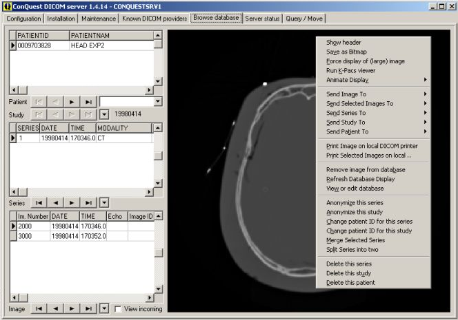
The Conquest DICOM project
Conquest is a full-featured DICOM server that handles DICOM-RT.
Marcel Van Herk and collaborators have developed and released
Conquest as an open-source package. The homepage is at http://ingenium.home.xs4all.nl/dicom.html.
Conquest is actively supported. Conquest has many
features,
including image format conversion, rules-based-forwarding, image
viewing, archiving, compression, archive merging, series merging,
etc.
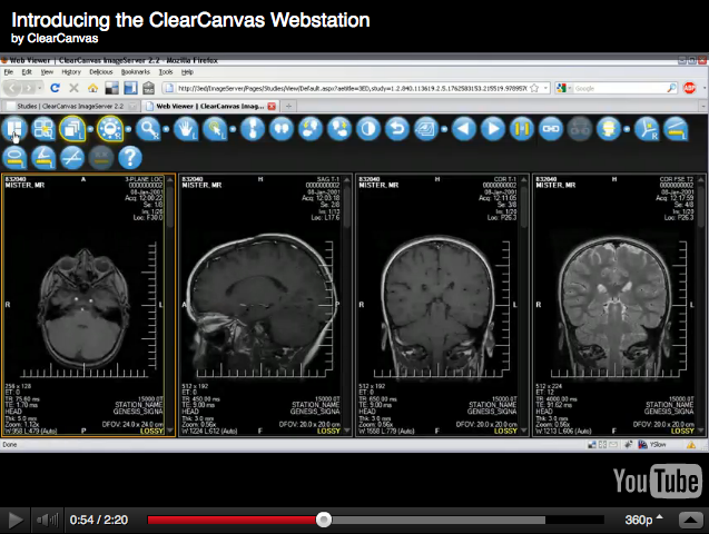
ClearCanvas
ClearCanvas is an open-source PACs server, embedded within an
application framework with a plug-in architecture. The
ClearCanvas viewer is entirely web-based (using the Microsoft
silverlight viewer), with server-side image processing. The
ClearCanvas
image server can be downloaded from http://www.clearcanvas.ca/dnn/Products/ImageServer/tabid/245/Default.aspx.
According to the website, ClearCanvas can handle some RT-specific
image
data, but does not handle RT-plan files (i.e., the DICOM component
with
the beam apertures, etc.).
Some interesting plug-ins that extend ClearCanvas functionality are
described at http://www.clearcanvas.ca/dnn/LiveDemo/tabid/204/Default.aspx.
Advanced plug-ins include tools for dynamic contrast enhanced
imaging
analysis.
Of note, ClearCanvas implements AIM, a standard for image
annotation.
The homepage of AIM is at https://cabig.nci.nih.gov/tools/AIM.
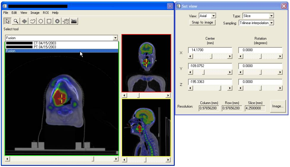
OSIRIX, an open-source DICOM PACs workstation and server
The home webpage is at http://www.osirix-viewer.com/.
However, at this time OSIRIX does not support RT-specific data
objects, such as dose distributions. Also, the FDA-cleared
version is (understandably) not free/open-source. An OSIRIX
app
is available for the iPhone/iPad. Here is a screenshot of the
FDA-cleared version:

Tools to anonymize radiotherapy
(DICOM-RT) data
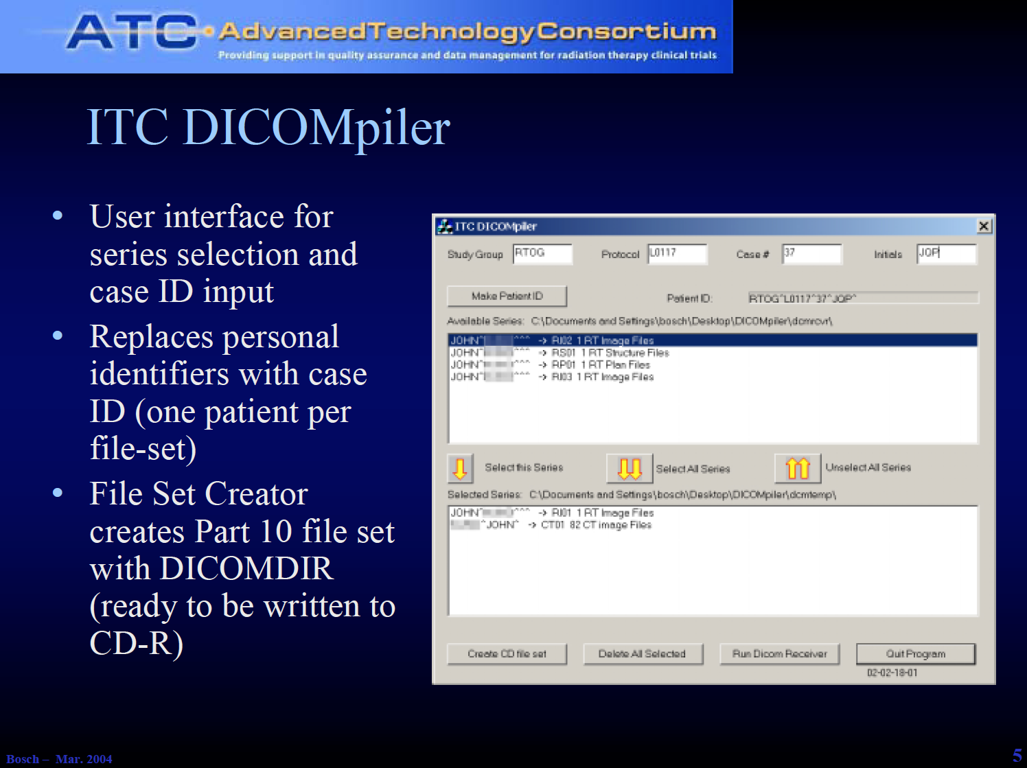
The DICOMPiler
The DICOMPiler, developed by the Image Guided Therapy Center at
Washington University in St. Louis, is a DICOM reciever that can be
used to conveniently anonymize DICOM series for subsequent
submission
in research protocols. The DICOMPiler can be found at http://itc.wustl.edu/DICOMpiler/index.htm.
Helpful instructions on the use of the DICOMPiler can be
found here.
CERR can also be used to anonymize data (see entry below.)
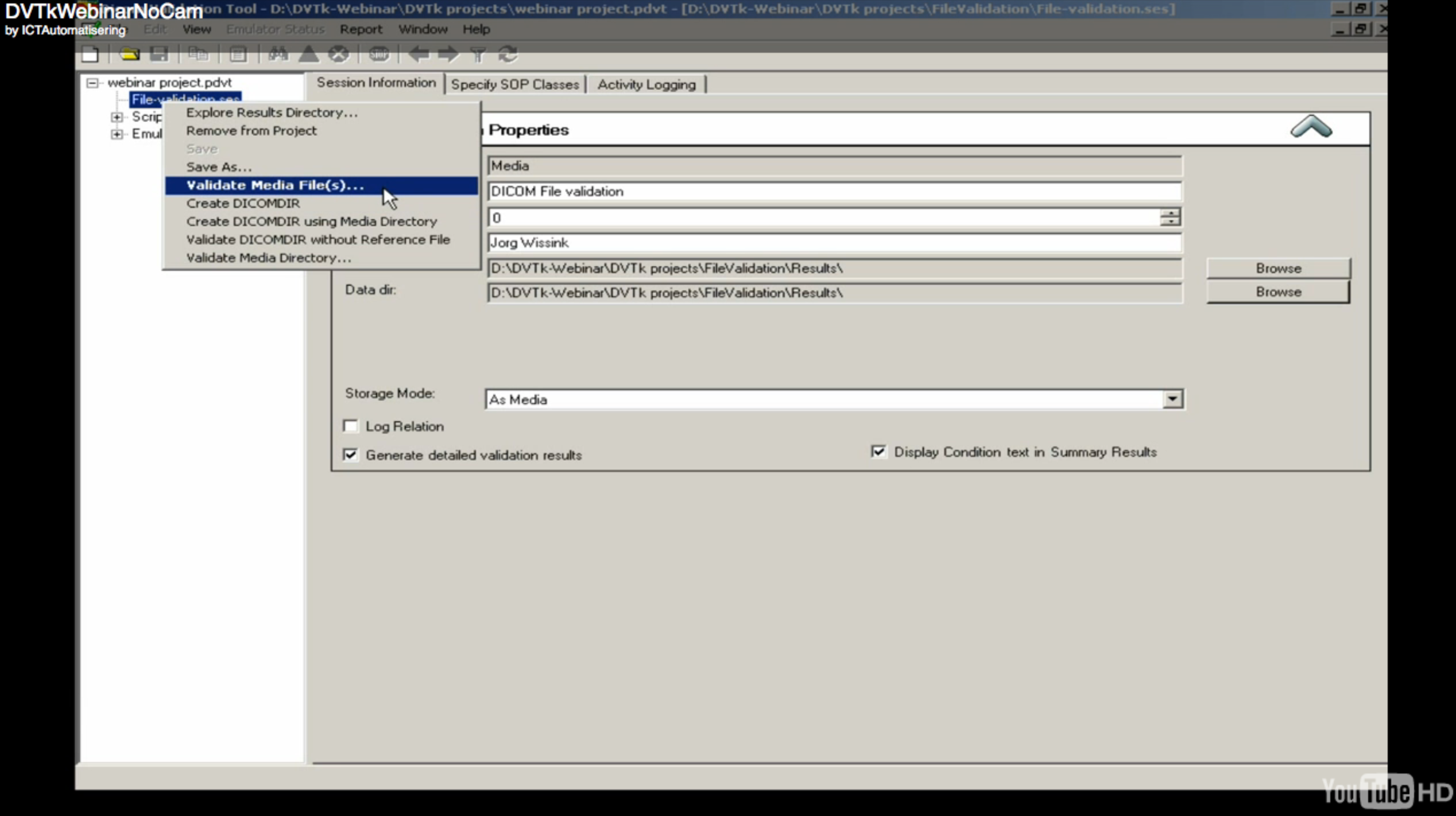
The DVTk project
DVTk is an open-source project to diagnose and validate DICOM
traffic
(DVTk: Dicom Validation Toolkit). It has further developed
high-quality DICOM-related applications, including an anonymizer,
editor, and a viewer. From the home page (http://dvtk.org): "DVTk is an
open
source project for testing, validating and diagnosing communication
protocols and scenario's in medical environments. It supports DICOM,
HL7 and IHE integration profiles."
Tools to manipulate radiotherapy
data
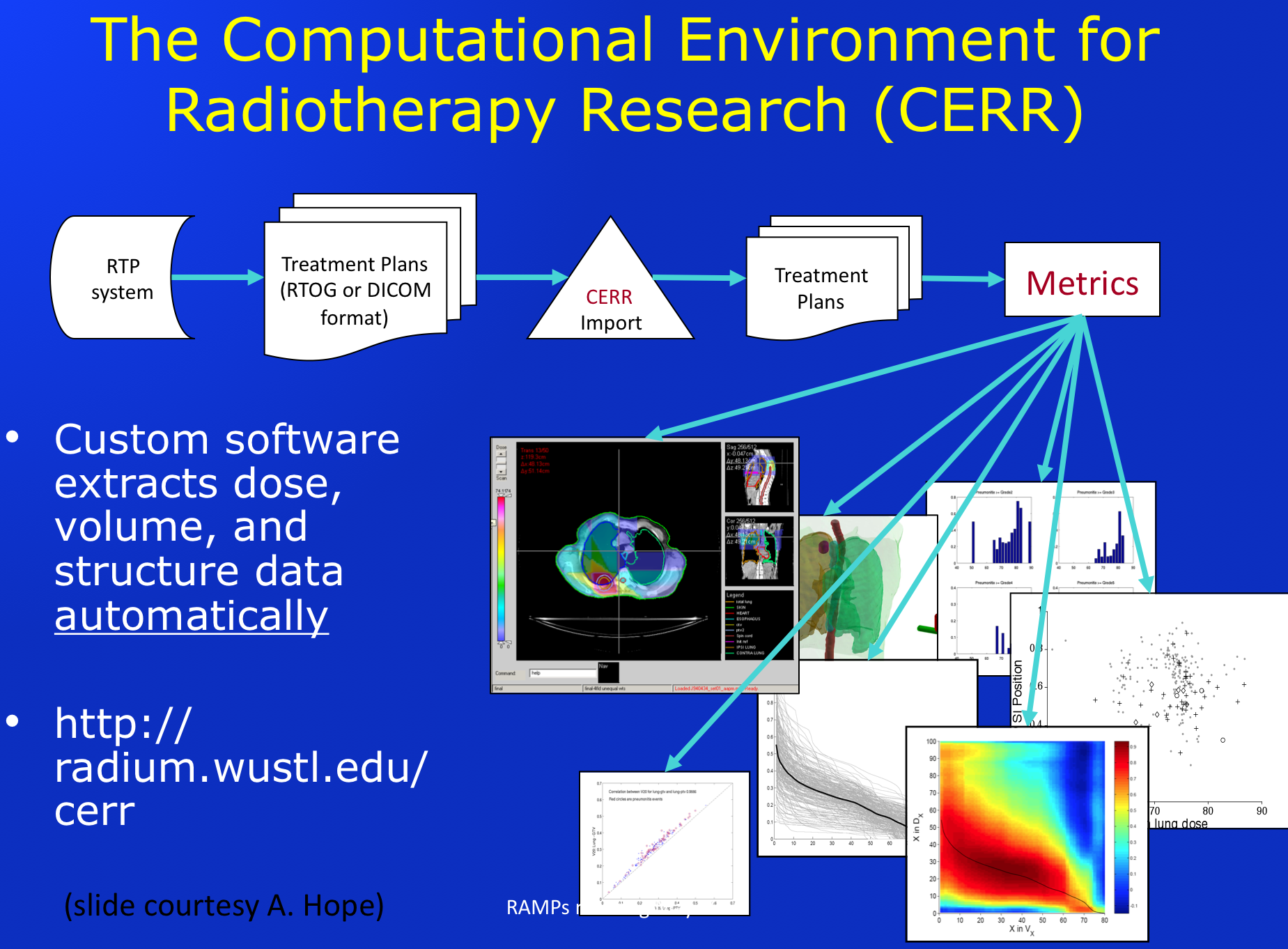
CERR: A Computational Environment for Radiotherapy
Research.
CERR is a widely used open-source system, based on the Matlab
platform,
that has comprehensive tools for manipulating radiotherapy data.
CERR has extensive import/export, visualization, image
registration, IMRT dose calculation, contouring,
complication/control
estimation tools, etc. The CERR homepage is at http://CERR.info. The CERR Wiki
is
at http://cerr.info/cerrwiki/index.php/CERR?w=CERRWiKi.
The dicompyler
Dicompyler is an ambitious open-source software system, based on the
widely used python language, that has many viewer and data analysis
features. In some ways, dicompyler parallels CERR. The
dicompyler homepage is at http://code.google.com/p/dicompyler/.
Dicompyler has a flexible plugin architecture, and a
convenient
graphical view of DICOM inputs.

RT_Image
RT_Image is an open-source software package developed by Edward
(Ted)
Graves, of Stanford University, that provides fairly general RT
image
viewing and data analysis tools. RT_Image can be downloaded
from http://rtimage.sourceforge.net/.
RT_Image has particularly strong PET-image viewing and
analysis
tools, including image contouring, registration, adn segmentation
tools, as it grew from those applications.
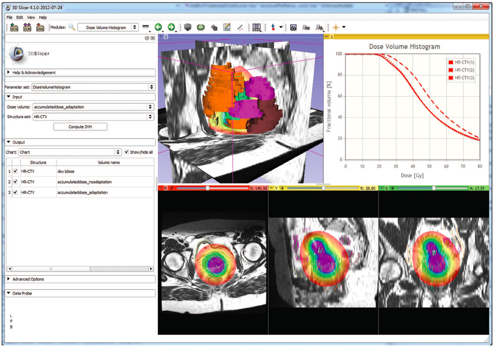
SlicerRT: an open-source tool for radiotherapy data
manipulation and processing built on Slicer.
Pinter, C., Lasso, A., Wang, A., Jaffray, D., & Fichtinger, G.
(2012). "SlicerRT: Radiation therapy research toolkit for 3D
Slicer". Medical physics, 39, 6332.
This paper was published just after the book chapter went to
print. Further information about this powerful package can be
found at
http://slicerrt.github.io/.
Here is the a screen shot from the paper by Pinter et al.:
Exporting/creating DICOM-RT structures
Gorthi S., Bach Cuadra M., Thiran J, "Exporting Contours to DICOM-RT
Structure Set," The Insight Journal - 2009 January - June.
http://hdl.handle.net/1926/1521. This paper and
associated
open-source code details methods for exporting structures in
DICOM-RT
format using ITK (see entry for ITK, below).
Medical Image viewing (general)
Slicer is an impressively comprehensive open-source, cross-platform,
software tool for visualizing and manipulating medical image data.
Some RT related capabilities have recently been added.
There is a dicomRT import capability, that imports structures
and
doses, based on the plastimatch software system. Slicer has
been
referenced in over 200 publications to date.
http://www.slicer.org/.
ITK, the Insight segmentation and registration ToolKit (ITK)
ITK is an extensive, stable, widely used open source toolkit
developed
by the National Library of Medicine to support image handling,
analysis, registration and segmentation. The ITK homepage can
be
found at http://www.itk.org/.
XiP: the eXtensible Imaging Platform and DICOM WG-23
Ca-Big has sponsored the development of XiP, the eXtensible Imaging
Platform. XiP is a platform of opensource tools to perform
rapid
prototyping of medical image processing and visualization
algorithms.
XiP utilizes an emerging protocol, DICOM WG-23 for
Application Hosting Interfaces, which has the goal of a
being a
widely adopted architecture for plugins. Thus, a researcher
who
writes a WG-23 compliant plugin could potentially transfer the
plugin
with little or no change to commercial systems that (hopefully)
adopy
the WG-23 standard.
Although XiP has great promise as a possible radiotherapy research
platform, RT specific data elements, such as dose distributions,
have
not yet been implemented.
The ca-BIG home page of XiP is https://cabig.nci.nih.gov/tools/XIP.
The wiki for XiP is http://www.openxip.org.
A powerpoint presentation on XiP that also describes WG-23 is given
at https://sites.google.com/a/barnett.id.au/interactive-workshop/dicom-wg23-1.
Slicer, a powerful opensource system for image viewing and
analysis.
The Slicer system is now widely used in various areas of image-based
medicine. The home page can be found at http://slicer.org. SlicerRT, a
package referenced above, was built using Slicer. Slicer is
deeply integrated with the ITK and VTK toolkits as well as many
community contributed extensions.
Image Processing (general)
ImageMagick
A particularly powerful image processing package is ImageMagick, the
result of an ongoing open source project, containing a large library
of
image manipulation command-line routines. ImageMagick can be
found here http://www.imagemagick.org/script/index.php.
imageJ
ImageJ is another powerful open-source image processing toolkit.
ImageJ, unlike imageMagick, can be used either as a library or
as
a stand-alone application. Many plug-ins have been developed
for
imageJ. The home page for imageJ is http://rsb.info.nih.gov/ij/.
Deformable Image Registration
Deformable image registration is a general technology, and many of
the
best tools are general.
Plastimatch (http://plastimatch.org)
is a deformable image registration system
developed by Greg Sharp and collaborators at Massachusetts General
Hospital/Harvard Medical School.
Statistical modeling (general)
The R-language
There are many open-source statistical modeling packages for
general use. The most well-developed suite of tools has been
developed around the R language, which was based on the Bell Labs
product, S. R contains a massive number of freely-available
routines, written by many thousands of contributors.
The wikipedia entry for S is at http://en.wikipedia.org/wiki/R_%28programming_language%29.
The official website of the R language is at http://www.r-project.org/.
Radiotherapy dose response
statistical
modeling
DREES: The Dose REsponse Explorer System.
Our group has developed a package to implement general model
selection
in the 'meso' modeling range (i.e. tens or hundreds of potential
predictor variables, but not thousands or millions).
DREES, the Dose-REsponse Explorer System, implements multivariable
logistic modeling building using cross validation techniques to test
the stability of model selection. It is open-source but
written
in the Matlab language. DREES can be found at http://CERR.info/DREES/.